Neutral red uptake (NRU assay) is one of the most widely used cytotoxicity and cell viability tests.
Cell viability tests are used to determine the proportion of viable cells after a cell maintenance procedure or to assess the cytotoxicity of a treatment.
In addition to the dye exclusion tests conducted with trypan blue, erythrosin B and other dyes, tests can be conducted in which you can assess the ability of live cells to retain a dye within the cytoplasm (dye uptake), as in the case of neutral red and dihydrofluorescein diacetate.
Furthermore, since the absorption of neutral red is proportional to the number of cells in culture, it is also possible to use the method to quantify cell growth and therefore, indirectly, the relative number of cells in culture.
The NRU test is recognized by the European Commission as a method to validate the acute toxicity of a molecule (see here) and by the OECD as a method to assess photocytotoxicity based on the relative reduction in viability of Balb / c 3T3 cells exposed to the chemical in the presence or absence of light (see here).
Neutral red
Neutral red is a relatively nontoxic supravital cationic dye.

Since neutral red has a pKa of 6.81, it is mainly in neutral form (NR) at physiological pH and can thus diffuse into the cell through the plasma membrane.
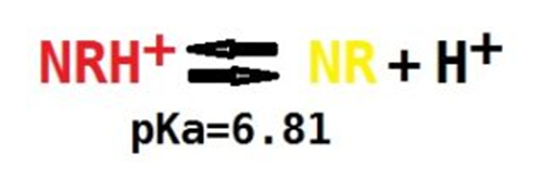
From the cytoplasm it spreads into intracellular organelles, remaining trapped in organelles such as lysosomes which, in functional and viable cells, have an acidic luminal pH. In fact, the dye become positively charged (cationic form, NRH+) at acidic pH and can no longer leave the lysosomes.
Since the acidification of lysosomes is an active process that requires consumption of ATP (accumulation of protons in lysosomes occurs through the active transport mechanism ATPase H +), the accumulation of neutral red in cationic form in the lysosomes will depend on the viability and metabolic state of the cell.
After exposure of the cells to the solution with the dye, the cells are washed and the dye extracted with a suitable extraction solution.
The absorbance of neutral red (proportional to the number of metabolically active cells with intact lysosomal membranes) is evaluated at a wavelength of 540 nm.
Reagents preparation
The preparation of all reagents can be done in advance, except for the neutral red working solution which must be prepared the day before the experiment.
PBS
see here for formulation, preparation and tips
NEUTRAL RED STOCK SOLUTION
Prepare a 4 mg / ml solution by dissolving 40 mg of neutral red in 10 ml of PBS.
Store the solution at room temperature in a dark glass container or covered with aluminum foil
NEUTRAL RED WORKING SOLUTION IN COLTURE MEDIUM
Working solution is a 40 μg/ml solution prepared fro the stock solution. In a sterile flask prepare a 1: 100 dilution of the stock solution in complete medium under aseptic conditions.
Mix 10 ul of stock solution + 990 ul of medium for a final volume of reaction mixture of 1 ml.
Prepare with this proportion the exact volume of reagent you need for the number of wells in which you want to test cell viability. This solution cannot be stored more then 1 day.
Incubate the mixture at 37 ° for 1 night.
The reaction mix must be prepared the day before the test is to be performed!
Before use it is then necessary to centrifuge the solution at 500 g for 10 minutes to remove the neutral red crystals
DYE EXCTRACTION SOLUTION
Prepare a solution with 50% ethanol and 1% glacial acetic acid
For 100 ml of dye exctraction solution put 49 ml of water, 50 ml of ethanol and 1 ml of glacial acetic acid in a bottle with a cap.
The solution can be prepared in advance and stored at room temperature in a tightly closed bottle
Protocol and Tips
The protocol can be carried out on cells plated in 96 or 24 multiwells.
In the first case the absorbance analysis must be carried out with a microplate reader, in the second case the analysis can be carried out with a microplate reader or with a spectrophotometer, if the first instrumentation -more and more recommended- is not available.
Protocol
- Prepare the cells.
Plate the cells in the wells at the desired density (you have to put enough cells for you to have a good reading, without going to saturation in the absorbance reading, see tip 1 to evaluate the concentration of cells to use) in culture medium.
| type of multiwell | 96 | 24 |
| ml of medium | 0,2 | 1 |
Generally, to avoid the edge effect in the wells on the perimeter of the multiwell the first and last row, as well as the first and last column are left for blank and filled with PBS or medium. These wells can be used as a blank, to test the possible interference of the culture medium with the neutral red
2. Let the cells grow for at least 24 hours, and treat them with the chemical or physical agent you want to test for cytotoxicity.


Here is a typical setup for experiments conducted in 96 or 24 multiwells. Clearly the 24 multiwell allow to evaluate the effects of a smaller number of concentrations, but more multiwells can be prepared. In this case it is always better to provide a blank column and one for the control even in the second multiwell.
For 96 multiwell, the first and last column as well as the first and last row are left empty (or better with PBS, to increase humidity and reduce evaporation in adjacent wells) to avoid the edge effect.
In the 24 multiwell the edge effect is not so marked and can be further reduced by placing sterile PBS in the spaces between the wells.
2. The day before starting the NRU test, prepare neutral red working solution in culture medium, as previously described and put it in the incubator for 24 hours.
3. Centrifuge the working solution at 600 g for 10 minutes at room temperature to remove any precipitated neutral red crystals.
4. Remove the medium from the wells and quickly rinse the wells with PBS.
| type of multiwell | 96 | 24 |
| ml of PBS | 0,5 | 1,5 |
Attention! this step can be avoided if the neutral red does not precipitate in the medium in the presence of the treatment or the carrier solvent to which the cells are exposed
Try to avoid this step especially in the presence of cells poorly adhering to the substrate.
5. Put the warm neutral red working solution in each well.
| type of multiwell | 96 | 24 |
| μl of working solution | 100 | 500 |
6. Incubate the cells for 1 to 2 hours in the incubator.
The shortest time should be chosen, sufficient to obtain a detectable signal, and generally 1 hour is sufficient.
The time can be increased up to 4 hours if cell density or metabolism is reduced.
7. At the end of the incubation time, observe the wells under a light microscope to evaluate the presence of precipitated crystals of neutral red.
If these are present, it is necessary to repeat the experiment (or discard the wells containing precipitates), for example by increasing the washing after treatment of the cells (some substances or solvents with which the cells are treated can favor the formation of precipitates) or by reducing the incubation time.
8. Carefully remove the neutral red solution
9. Wash each well with PBS twice
| type of multiwell | 96 | 24 |
| μl of PBS | 200 | 1000 |
10. add the destain solution to each well
| type of multiwell | 96 | 24 |
| μl of destain solution | 150 | 800 |
11. Let the neutral red dissolve by placing the multiwell plate on an orbital shaker for 20 minutes.
12. Measure absorbance at 540 nm with microplate reader
For greater sensitivity and less interference it would be optimal to use a light with excitation and emission wavelengths at 530 and 645 nm respectively.
Analysis and interpretation of the results
CYTOTOXICITY TESTS
Citotoxicity is calculated by comparing the absorbance of the sample treated with the molecule to be tested to that of the control.
1) subtract the absorbance values of the blank from the absorbance values recorded
Areal = A-Ab
2) calculate the relative intensity of absorbance in the treated versus that of the control
Ar = At / Ac
3) create a Cartesian diagram the concentration of the molecule with which the treatment was carried out on the abscissa and the relative absorbance on the ordinate
4) calculate the LC50 by fitting as effectively explained in this video
This article contains some important considerations for the calculation
Tip 1
EVALUATION OF THE TEST LINEARITY
Since the response of the absorbance as a function of the concentration is linear only within a certain range of values, it will be necessary to first construct a calibration line for the number of cells / absorbance to define which is the range of cell density to plate so that the absorbance of the neutral red falls in the linear portion of the calibration line.
In general with this test a good linearity of response is guaranteed (absorbance values lower than 1.2 even in the presence of confluent cells).
However, as this depends very much on the type of cells and the culture conditions, it is advisable to repeat this calibration test every time this test is used for the first time with a new cell line.
To do this, it is necessary to evaluate under control conditions the absorbance of the neutral red released after treatment of cells plated at different concentrations.