Erythrosin B can be used for cell viability test.
Cell viability is an indicator of the overall health and integrity of cells within a sample.
Before carrying out any cell experiments aimed at characterizing morphology, pathophysiological responses, and so on, you need to be certain that the cells in your culture are viable.
Furthermore, cell viability may be of interest after exposure of the cultured cells to an agent whose cytotoxic effect is to be determined (chemotherapy drugs) and/or excluded (cosmetics or food additives and so on).
Viability is often defined as the number, or rather the percentage, of healthy cells in a sample’s overall cell population.
Nowadays, several viability tests are used, but the widely used dye exclusion test remains one of the simplest and fastest to perform.
Contents
- Erythrosin B
- Protocol
- Erythrosin B viability test: semi-authomatic method with Fiji
- Final considerations and limitations
Erythrosin B
Erythrosin B is a tetraiodofluorescein dye commonly used as a food and drug additive, but also widely applied in cell biology.
Its principle of action is straightforward: Like trypan blue,
Viable cells exclude the dye thanks to their intact membranes.
Non-viable cells absorb the dye and appear pink to red under the microscope.
Why Use Erythrosin B Instead of Trypan Blue?
Using erythrosin B instead of trypan blue offers several advantages:
- not toxic (for cells – even after 2 hours of incubation- and for users),
- it does not bind to serum proteins,
- not require an incubation period before counting (1-2 minutes is enough)
The erythrosin solution used for the exclusion test contains the dye at a concentration of 0.1 or 0.2% (2 mg in 1 ml of solution)
and, like trypan, it must be mixed in a 1: 1 ratio with the cell suspension whose viability must be determined.
By not binding to serum proteins, the contrast between the stained cells and the background is better than trypan even when leaving the cells in the medium during counting.
Non-viable cells take on a red (pink) color.
The drawbacks of erythrosin are
- Photosensitivity and hygroscopicity.
Keep the container tightly closed in a dry and well-ventilated place!! Keep the container away from light
- Incompatibility with oxidizing agents.
Traditionally this test involves the cell count (with distinction between live and dead cells) with a hemacytometer viewed under an optical microscope (manual method).
Nowadays, semi-automatic method can be used (with image acquisition and use of Fiji, see what’s written for trypan blue) or totally automatic with the use of coulter counters that provide quick results (Soo In et al, 2016).
Erythrosin B has also been used successfully as a vital dye for monolayer cells (Krause et al., 1984)
Protocol
PREPARATION OF ERYTHROSIN B 2% STOCK SOLUTION IN PBS
- weigh 0.2 g of erythrosine in a falcon and add 10 ml of PBS at pH 7.4
- vortex until completely dissolved
- filter with a 0.45 um filter
- aliquot in 1 ml aliquots in dark glass containers or eppederf wrapped with aluminum foil.
The stock solution is stable at 4 °C and should be stored protected from light. Using PBS helps maintain pH and prolongs shelf life.
At neutral pH erythrosine is soluble, while at acid or basic pH it forms precipitates (yellow or red respectively).
PREPARATION OF ERYTHROSIN B 0.1% WORKING SOLUTION IN MEDIUM
Vortex erythrosin B stock solution until homogenous.
Prepare a 0.1% solution of by pipetting 50 ul of stock sulution into 950ul of complete culture medium. Medium is used instead of PBS to avoid stressing the cells during the viability test, particularly in live-cell protocols.
Vortex erythrosin B working solution until homogenous.
Since the dye is photosensitive, the solution must be shielded by wrapping the container in a piece of aluminum foil or placing it in a dark glass bottle.
CELL COUNT
A. After treatment with the substance of interest,
- if the cells are adherent to the growth vessel, you must detach them by the method you usually use (trypsinization or any other methods you use when you have to subculture the cells)
- if cells grow in suspension, centrifuge to pellet
TIP 1 FOR ADHERENT CELLS
Note that during trypsinization most of the non-viable cells will be lost in the PBS during the first wash to remove the serum, prior to trypsin treatment.
For an accurate assessment of the number of non-viable cells, it will therefore be necessary to recover the wash PBS by combining it (in the same eppendorf) with the trypsinized cell suspension and pelleting the cells deriving from washing and trypsinization for the viability test.
Trypsin is a rather harsh treatment for detaching cells and could further damage cells upon exposure to a cytotoxic agent. For this reason it is better to choose the gentlest possible subculture method that may not be trypsin
B. resuspend the pelletted cells in the growth medium so that the cell suspension have a cell concentration between 2×10⁶ and 106 cells / ml
TIP 2
This value of cell concentration is chosen in order to have a good number of cells (between 100 and 50) subsequently in the hemocytometer grid.
It is an indicative value that allows you to have not too many and not too few cells to count, in order to minimize the error having reasonable accuracy and precision.
Furthermore, for the subsequent count it is imperative that the cells are well separated (avoid cell clumping) from each other.
C. Make a 1: 1 dilution of the cell suspension with 0.1% erythrosin B solution and smoothly mix.
In practice, after mixing the cell solution carefully and thoroughly (remember that the cells settle very quickly!) take 50 µl of the cell suspension, put them in an eppendorf and add an equal volume (50 µl ) of erythrosin B solution. Avoid foaming
TIP 3
The 50 µl volume of cell suspension and trypan is only indicative, you can choose a desired volume. However, the volume must not be too small so that a deep mixing between the solutions is guaranteed, possibly avoiding the formation of foam!
D. Prepare the hemocytometer and count the cells as indicated here
E. Count cells in at least 4 quadrants, distinguishing pink (dead) from unstained (viable) cells. Take an average of the value obtained
TIP 4
Since you have made a dilution of the initial cell suspension with equal volume of erithrosin B, it will be necessary to multiply the value of cells / ml by 2 to get the total number of cells in the cell suspension.
F. Calculate the% of viable and dead cells
% viable cells = (number of unstained cells / total number of cells)x100
% dead cells = (number of stained cells / total number of cells)x 100
G. Repeat the counting procedure at least two more times by repeating steps D to F
Erythrosin B viability test: semi-authomatic method with Fiji
This method is completely analogous, in the cell preparation phase, to the previous one.
However, it does require image capture with a micro camera.
Advantages:
- you can save images and results for future documentation
- It allows to increase the speed of the counts
- It also improves the accuracy of the count performed by using an analysis program after acquisition (a posteriori).
Fiji is a totally free program that can be downloaded here. Fiji is an image processing package with a lot of plugins that facilitate image analysis. Here we will use the Cell count plugin
Protocol
Proceed as above from A to C.
D. load the cell suspension into the hemocytometer, focus and acquire the image (or images relating to the different treatments whose cytotoxicity is to be evaluated).
TIP 1
Since you carry out the cell count a posteriori, you can to prepare a certain number of samples at the same time for rapid acquisition one after the other..
E. Open Fiji
F. Open the first image you captured (File –> Open)
G. Activate the Plugin Cell Counter (Plugins –>Analyze–>Cell Counter). Flag on “Keep original”,and click on Initilize. A duplicate of your image will be created.
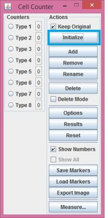
H. Select the Type 1 counter and click Rename. A dilaog box will appear where you can rename your counter Type, let’s call it “alive” for example
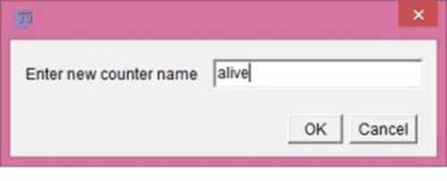
I. Rename also the Type 2 counter with “dead”
J. Now select the alive counter, go on the original image and click on each of the alive cell that you can see in your image (number 1 on the image). Then select the “dead” counter and do the same with the blue coloured cells. You’ll end up with all the cells selected.
K. Click on “result”. So that you’ll end up with a table showing the number of dead cells and the number of alive cells
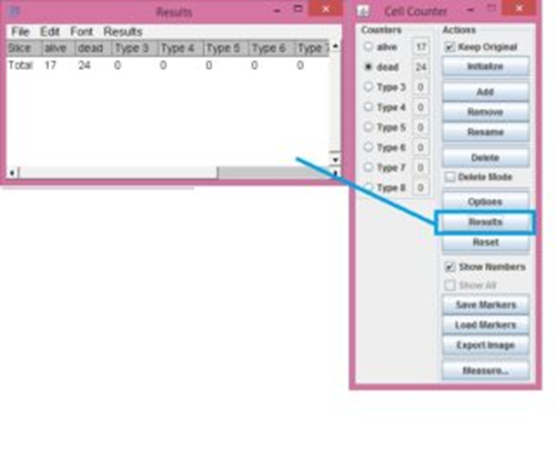
L. Calculate the % of viable cells
%viable cells=[Number of alive cells/(number of alive cells+number of dead cells)]x100
Final considerations and limitations
In addition to erythrosin B, other dyes such as naphthalene black, eosin, Congo red or propidium iodide can be used in the dye exclusion test.
While performing the dye exclusion test, some limitations must be kept in mind
- irreversibly damaged cells can take several days to lose the integrity of their membrane, so the damage would not be detected with this type of test
- some damaged cells may undergo premature disintegration so they are not detected in the dead cell count.
These factors can cause cell death to be underestimated.