Why is phenol red added to culture media?
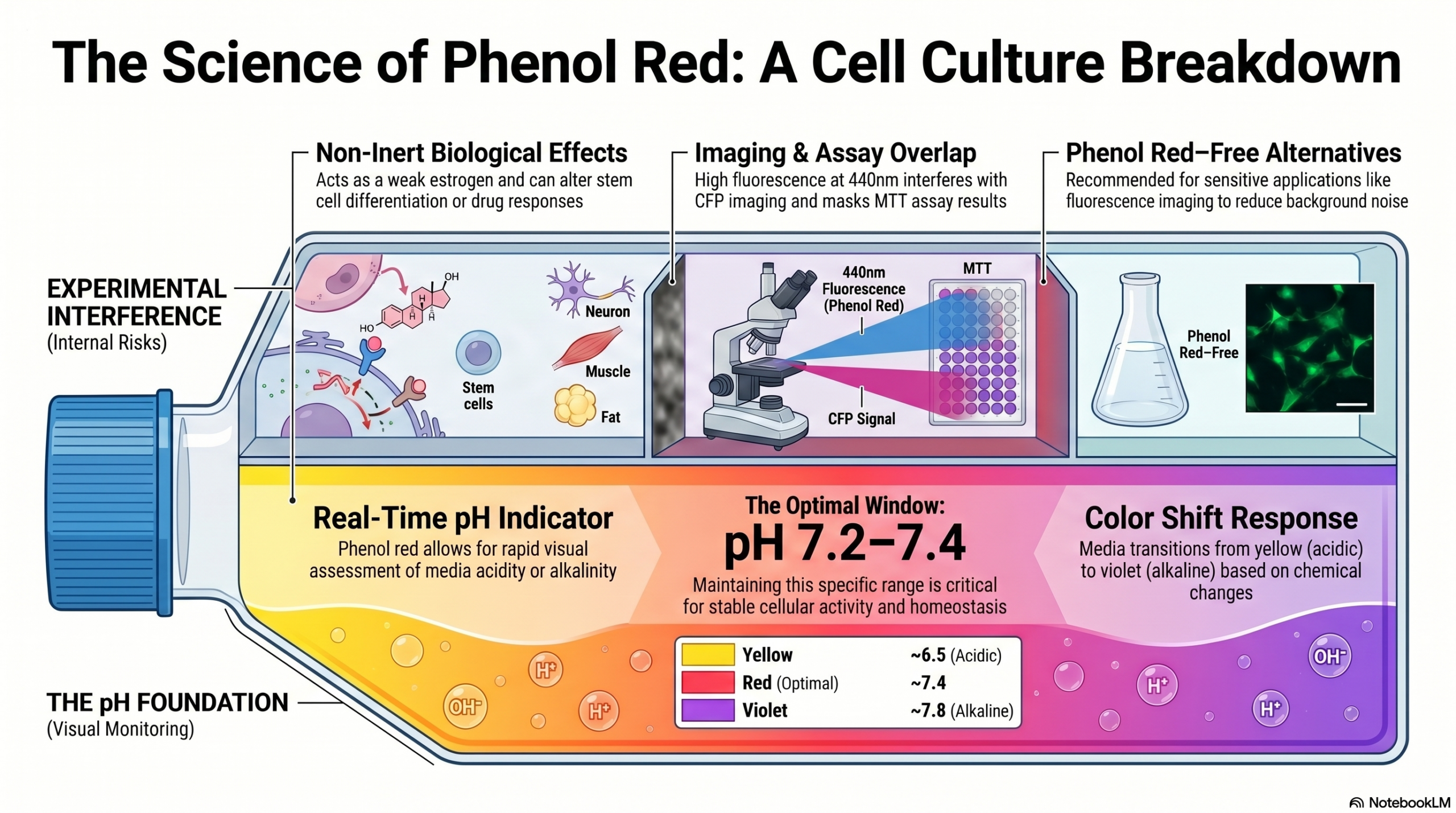
Phenol red is commonly added to cell culture media as a pH indicator.
It changes color depending on pH:
- red at pH ~7.4
- orange at pH ~7.0
- yellow at pH ~6.5
- pink at pH ~7.6
- violet at pH ~7.8
This allows a rapid visual assessment of the pH of the culture medium.
The pH of the medium should be maintained around 7.2–7.4, as pH strongly affects cellular activity.
Cells and organisms have multiple mechanisms to maintain pH homeostasis, highlighting the importance of this parameter.
Phenol red concentration varies depending on the medium formulation, but it is typically present in the range of 5-15 mg/mL .
Be careful: is phenol red really inert?
Phenol red is often considered an inert compound.
However several studies suggest that this indicator can affect cellular activity to varying degrees.
🔬 Reported biological effects
Phenol red has been described as:
- a weak estrogenic compound under certain conditions (Y Berthois et al. 1986, Welshons WV et al., 1988, Faria et al., 2016, Liu et al., 2013, Lyshdal et al., 2013, Wesierska-Gadek J et al., 2007, Still et al. 2003)
- capable of upregulating CFTR expression in vitro (Tsang et al., 2001)
- able to mask or alter drug responses (e.g. roscovitine)
- affect chondrogenic and osteogenic differentiation of mesenchymal stem cells (Lysdahl H et al.)
- regulate keratin expression in keratinocytes (Aldehlawi et al., 2020)
- exhibit redox activity (Morgan et al., 2019).
👉 These effects highlight the need for careful evaluation when using phenol red in experimental design.
Interference with colorimetric assays
Before performing colorimetric assays, it is necessary to assess whether there is spectral overlap between assay reagents and phenol red.
For example significant overlap exists between phenol red and MTT assay reagents.
Possible solutions:
- Use phenol red–free medium
- Remove the medium and dissolve formazan crystals in DMSO
- Include appropriate background controls (medium + reagent, without cells)
Important:
The color (and absorbance) of phenol red depends on pH.
👉 If the medium becomes acidified by treatment or increased cell density, correction methods may introduce errors. More advanced correction methods (e.g. acidification with HCl) can be used when necessary.
Interference with fluorescence imaging
Phenol red is highly fluorescent when excited at around 440 nm.
👉 It interferes with imaging of cyan fluorescent proteins (CFP). Phenol red free media has to be used.
Additionally phenol red increases background fluorescence, reducing image quality. For fluorescence imaging, phenol red–free media are recommended (see here for more tips on media for fluorescence imaging, or here for an example and discussion).
Other experimental uses of phenol red
Phenol red is also used in various research applications, including:
- assays for superoxide and hydrogen peroxide production by macrophages (1, 2)
- peroxidase activity tests, such as vanadium bromoperoxidase (3, 4)
- detection of eosinophils and eosinophilic peroxidase in situ (5)
- detection of halogens (e.g. bromide, iodide) (6, 7)
- potential applications in cancer cell detection (8)
Conclusion
Phenol red is a useful and widely used pH indicator that enables rapid monitoring of culture conditions.
However, it is not biologically inert and can influence cellular processes, assay results, and imaging quality.
👉 Its use should therefore be carefully evaluated depending on the experimental context.