
BACKGROUND
The amount of proteins (and, therefore, indirectly, of cells) in a sample can be quantified by directly evaluating the absorbance at 280 nm. The relationship of absorbance at 280 nm to protein concentration is linear.
The aromatic rings of several aminoacids (mainly tryptophan and tyrosine and to a lesser extent to phenialanine) of the proteins in solution absorb ultraviolet light at a wavelength of 280 nm.
Absorbance at 280 nm can be used to evaluate a minimum of 100 μg of proteins (at a concentration ranging from 20 to 3000 ug/ml) – which corresponds roughly to 200,000 cells.
PROS AND CONS
This is a quick and convenient method, as no additional reagents or incubations are required.
However, all non-protein component of the solution that absorbs ultraviolet light at 280 nm could interfere with the measurements. Since different proteins, nucleic acids and intracellular metabolite have very variable absorption characteristics, there can be considerable error, especially in complex mixtures such as cell lysates.
The main interfering substances in addition to nucleic acids, nucleotides such as ATP or NAD(P), heme-containing compounds such as cytochromes, reagents containing sulfhydryl groups such as 2-mercaptoethanol, dithiotreitol, glutathione or cysteine.
The absorption spectrum of tyrosine is pH dependent, but at 280 nm both the protonated and non-protonated forms have similar absorption coefficients and therefore it is not necessary to perform measurements at a particular pH.
The absorption spectrum of tyrosine depends on pH, but the protonated and non-protonated forms of this amino acid have similar absorption coefficients at 280 nm and therefore it is not necessary to perform measurements at a particular pH. However, as the secondary, tertiary and quaternary structure of proteins can influence absorbance, factors such as pH, ionic strength, etc. must be kept constant in the different samples under examination.
Be careful if you use DTT in protein solubilization buffer! DTT oxidizes over time leaving a product that absorbs at 280 nm. If you freeze the protein samples and then thaw the sample after a certain period of time, it is not possible to know the concentration of the oxidized DTT and this can affect the measurement!
As glass and plastic absorb in UV it is necessary to use quartz cuvettes (but there are on the market disposable UV transparent plastic cuvettes as well)
PROTOCOL (WARBURG AND CHRISTIAN METHOD)
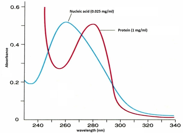
Figure 1. Absorption spectrum for nucleic acids and proteins. Nucleic acids have a peak absorption at 260 nm, proteins at 280 nm. However, nucleic acids also absorb light radiation at 280 nm, for this reason in the dosage of proteins in a cellular lysate their presence and interference must be taken into account
An important note before starting: Since we are working with a raw lysate of cells, which, in addition to proteins, contains nucleic acids, it is necessary to use a method (Warburg and Christian) that takes into account the interference by the nitrogenous bases of nucleic acids that also absorb (albeit less efficiently) electromagnetic radiation at 280 nm (figure 1).
- Dissolve Cell Monolayers and / or Cell Pellets in an appropriate volume lysis buffer (as an exdample: 0.5-1.0 M NaOH by heating them to 100 ° C for 30 minutes or by leaving them overnight at room temperature. Alternatively, you can use a solution with 0.3 M NaOH and 1% sodium lauryl sulfate (SLS). In this case you have to wait 30 min at room temperature).
- To avoid light scattering, it is important to check that the protein solution is not turbid. Centrifuge at 10000 rpm for 10 minutes at 4 °C to remove cell debries.
- Turn on the UV lamp of the spectrophotometer (or plate reader)
- Select absorbance reading at 280 nm
- Correct for the background with a cuvette containing only the solution in which you lyzed the cells
- Measure absorbance of the protein solution and subtract that of the blank
To evaluate nucleic acid interference
- Select absorbance reading at 260 nm
- Calibrate to zero absorbance with buffer solution only
- Measure absorbance of the protein solution
To evaluate the presence of cellular debris that increase the turbidity of the solution, the absorbance can be checked at 320 which should be less than 0.02 – see below.
BE CAREFUL
Cold solutions can fog up the cuvette, while warm solutions can form bubbles and interfere with the readings.
For concentrated solutions (absorbance greater than 1.4) simply dilute the solution.
CALCULATION
Use the following formula to estimate the protein concentration of a cell lysate:
Concentration (mg/ml) = [(1.55 x A280) – (0.76 x A260)]*DF
where A280 is the absorbance measured at 280 nm
A260 is the absorbance measured at 260 nm (nucleic acid correction)
and DF is the diluction fator express how much you diluted your protein. If you’ve your starting protein solution the dilutio factor is 1, if you dilute your protein (becuse the absorbance excess 1.5) in 1:2 ratio, then the dilution factor is 2, and so on.
This formula, by Warburg and Christian, is for a 1cm pathlength. The absorbance at 260 nm (A260) accounts for nucleic acid interference. (Warburg, O. and Christian, W. Biochem. Z. 310: 384 (1941)).
You can use an additional background correction (A280-A320) that takes into account the presence of the dispersion of particles in solution (such as cell debris). Anyway, the values for A320 must be less than 0.02; otherwise, there is a significant amounts of impurities in the sample.
To convert units, use these relationships:
% of protein = mg protein /ml divided by 10
in fact if for example you have a 1 mg / ml solution 1mg / ml = 0.001g / ml which is equal to 0.1g in 100ml, i.e. the solution is 0.1%.